FREE Consultation

During this 30 min appointment, we will perform a basic eye assessment to determine whether you are suitable for refractive surgery.

The needs of each patient dictate the way we employ our Laser Eye Surgery technology.

Some Laser Eye Surgery clinics still work with relatively basic information and measurements, such as your lens prescription and wavefront analysis. But at London Vision Clinic, we know that world-class Laser Eye Surgery treatment requires much more.

We understand that no two eyes are the same. So, when considering prospective patients, our first task is to make sure we understand your eyes completely, down to the finest details. To do this, we employ the best possible technology available. Our personalised treatment plans are based on the results of our comprehensive screening process and years of pioneering ophthalmic research. This ensures your treatment is tailored specifically to each of your unique eyes.

Our laser eye surgeons don’t just train with specialist technology! They help design it.

Artemis Insight 100™ scanners. Deemed the most accurate corneal scanner in the world, this impressive technology was developed by our very own founder and expert surgeon, Professor Dan Reinstein. Since its invention during the 1990s, the Artmeis Insight 100™ has greatly enhanced patient safety and refractive outcomes across the Laser Eye Surgery industry.

The superior imaging and biometric capabilities of this technology allow us to confidently diagnose eye diseases earlier and treat patients who might previously have been rejected for Laser Eye Surgery.

London Vision Clinic was the first in the world to have the Artemis Insight

London Vision Clinic’s unique partnership with Carl Zeiss Meditec, a world-leading laser manufacturer.

Our Laser Technology

MEL 90 Excimer laser

Carl Zeiss Meditec
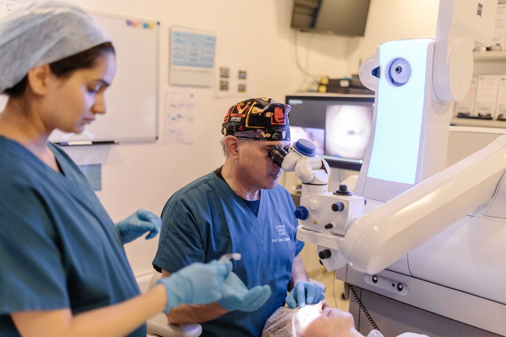
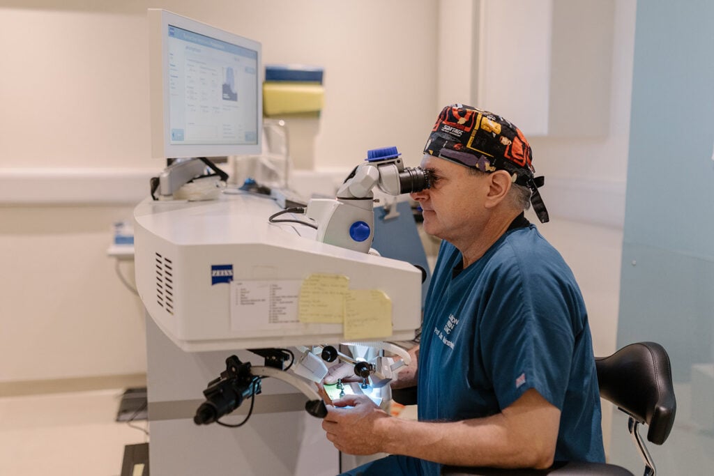
Ophthalmologist in blue scrubs uses a surgical microscope at a workstation with a computer monitor nearby in a clinical setting.

The Carl Zeiss Meditec MEL 90 excimer laser offers unrivalled accuracy in its auto-stabilised, calibrated laser energy delivery. It uses closed-loop energy stabilisation, the first laser to do so.

This gives the fastest response time of any laser in the world without the need for frequent user calibration. 

Professor Reinstein was part of the Carl Zeiss Meditec team that developed the MEL 80 and MEL 90 lasers. These state-of-the-art lasers have been proven to be the most accurate refractive lasers in the world, as determined by rigorous US FDA trials.

VISUMAX 800 Femtosecond laser

Carl Zeiss Meditec
VISUMAX800

The Carl Zeiss Meditec VISUMAX 800 is the latest-generation laser used to perform SMILE Pro keyhole laser eye surgery. More than 12 million SMILE procedures have been performed worldwide—far more than with any other type of lenticule-extraction laser—providing an unrivalled depth of experience with the technology.

The VISUMAX 800 completes the laser treatment in less than 10 seconds, around three times faster than the previous generation. It also helps the surgeon position the treatment accurately and account for any rotation of the eye, which is particularly important when correcting astigmatism. The eye is held steady using gentle suction, so patients usually feel only mild pressure. This also reduces the likelihood of small red marks developing on the white of the eye that are common with lasers that use higher pressure.

Imaging And Diagnostics Devices

Artemis Insight 100™ Ultrasound Scanner

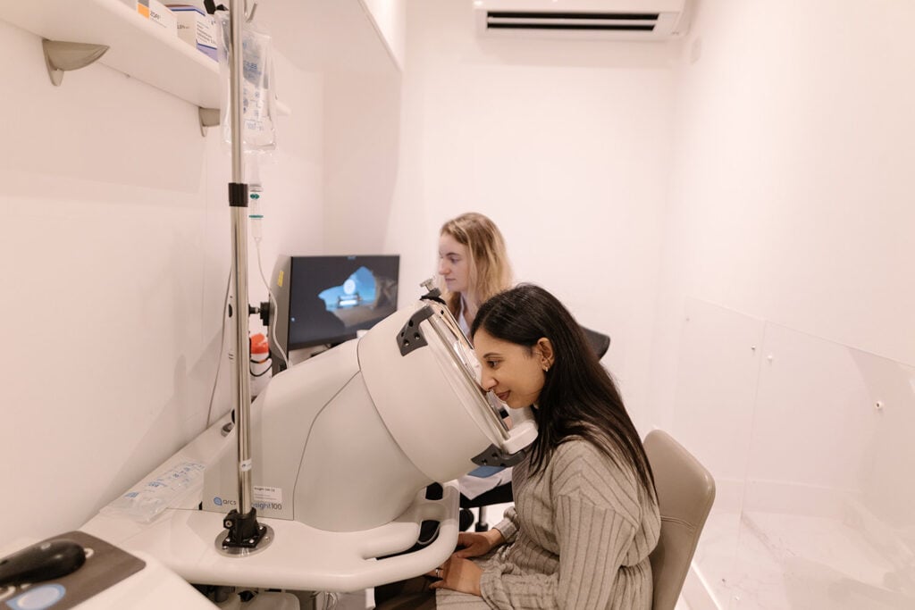
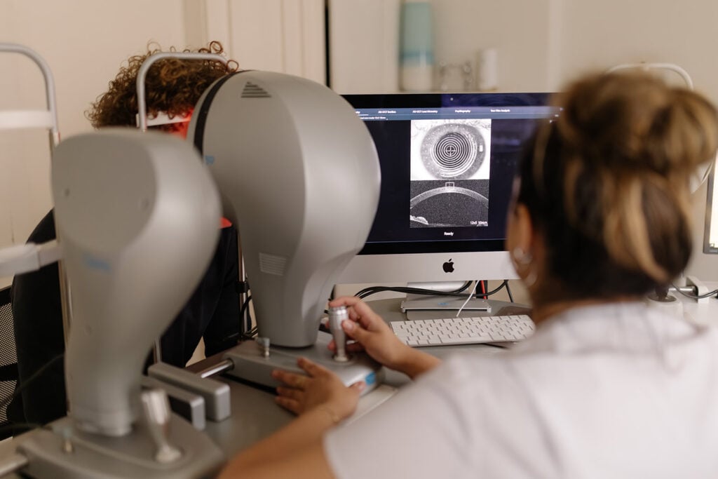
Patient sits at an ophthalmology device with chin on rest as a clinician watches a monitor displaying an eye image.

The Artemis Insight 100™ Ultrasound Scanner is the world’s most accurate corneal scanner. It provides precise measurements for epithelial and corneal thickness for Laser Eye Surgery and accurate measurements from behind the iris for ICL surgery. 

Accurate to 1 micron (1/1000th of a millimetre) in the cornea, it is the most accurate method for planning the anatomy of any vision correction surgery. This pioneering technology resulted from bio-engineering ophthalmic research conducted by Professor Reinstein and his coworkers at Cornell University in New York in the early 1990s.

The Artemis Insight™ is more accurate than optical coherence tomography (OCT), tomography or hand-held ultrasound machines. As such, it is the most sensitive method of mapping the individual layers of the cornea, as well as the internal structures of the eye. This makes it invaluable in the planning of Laser Eye Surgery, ICL surgery, and cataract surgery.
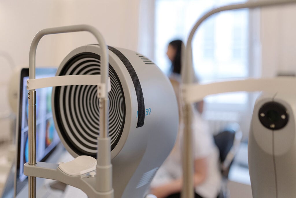
CSO MS39 Optical Coherence Tomographer (OCT)

MS39-OCT

Produces highly detailed maps of the cornea and its individual layers, supporting accurate screening and planning for laser eye surgery and lens procedures.

Oculus Pentacam HR Tomographer

Creates a detailed 3D map of the cornea and front of the eye, supporting accurate screening and planning for laser eye surgery or lens procedures.

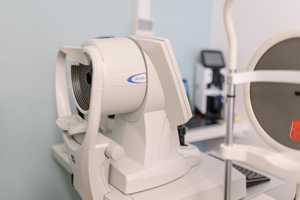
ZEISS Atlas 9000 Corneal Topographer

White Atlas medical imaging device in a clinical room, ready for use by a technician.

Creates a detailed map of the cornea, helping assess eye shape, detect irregularities and plan personalised laser eye surgery with exceptional precision.

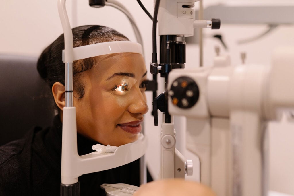
Topcon SL-D7 Digital Slit-Lamp

hyperopia-treatment-london

Used by the clinicians to perform a detailed examination of the front, internal, and back of the eye, helping assess eye health before and after laser or lens surgery.

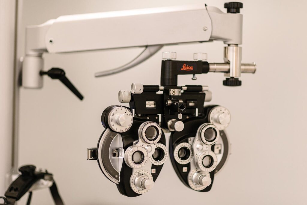
Leica Manual Phoropter

Leica phoropter used for eye exams, with multiple dials and lenses mounted on an arm.

Used during your eye examination to accurately measure your prescription and determine the clearest, most comfortable vision before laser eye surgery or lens replacement procedures.

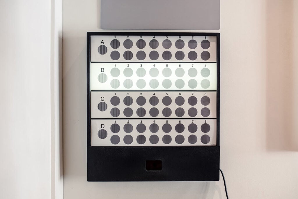
VectorVision CSV-1000

Close-up of a grid/chart with gray circular markers in rows A and B and columns 1–4 on a display panel.

Measures visual sharpness and contrast sensitivity under different lighting conditions, helping assess real-world vision quality before and after eye surgery.

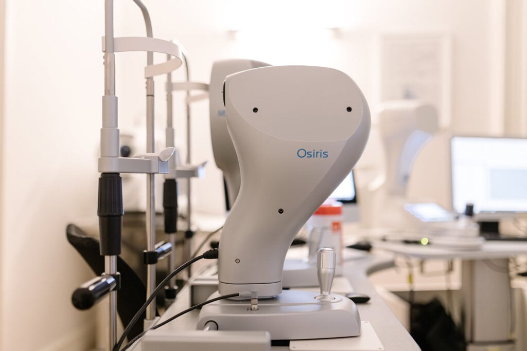
CSO Osiris Aberrometer

White Osiris robotic arm on a lab bench with monitors and equipment in the background.

Measures subtle optical imperfections across the entire eye, helping guide personalised treatment planning and assess visual quality before and after eye surgery.

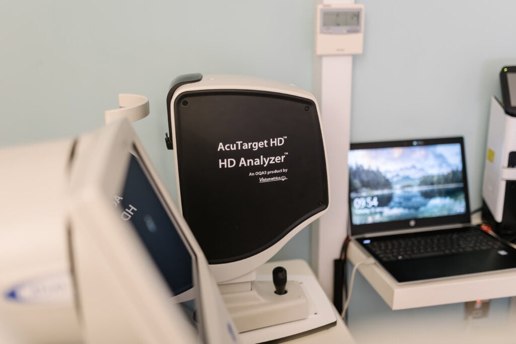
HD Analyzer

HD Analyzer

Measures the eye’s optical quality, light scatter and tear-film stability, helping assess visual performance before and after laser eye surgery or lens procedures.

MS-39 Dynamic Pupillometry

MS39 dynamic pupillometry

Tracks how the pupils respond under different lighting conditions, helping personalise treatment planning and assess potential night-vision symptoms before eye surgery.

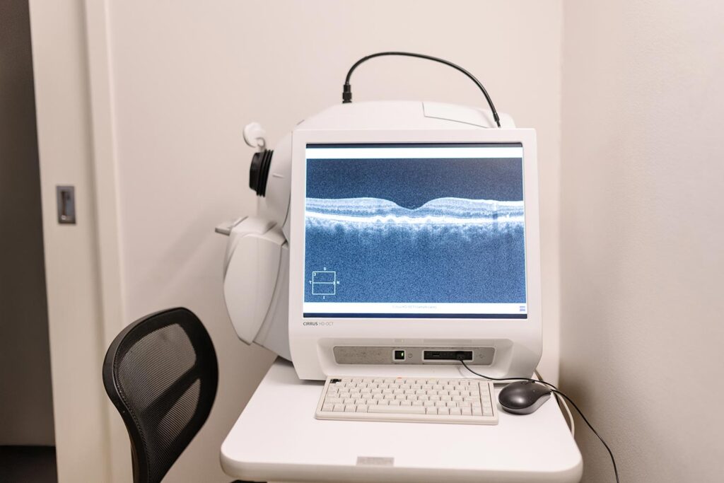
ZEISS CIRRUS HD-OCT Scanner

Optical coherence tomography (OCT) scanner on a desk with a monitor displaying a retinal cross-section image, in a clinic room.

Creates high-resolution cross-sectional images of the retina and optic nerve to assess eye health before laser eye surgery or lens surgery, and monitor long-term outcomes after treatment.

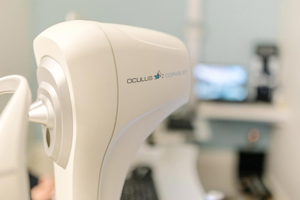
OCULUS Corvis® ST

oculus

Measures the biomechanical strength and response of the cornea, helping assess suitability for laser eye surgery and supporting personalised treatment planning.

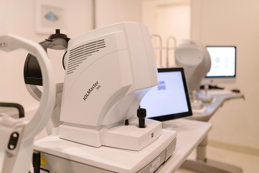
ZEISS IOLMaster® 700 Biometry

Uses advanced optical biometry to measure the eye with exceptional precision, helping select the most accurate lens implant for cataract and lens replacement surgery.

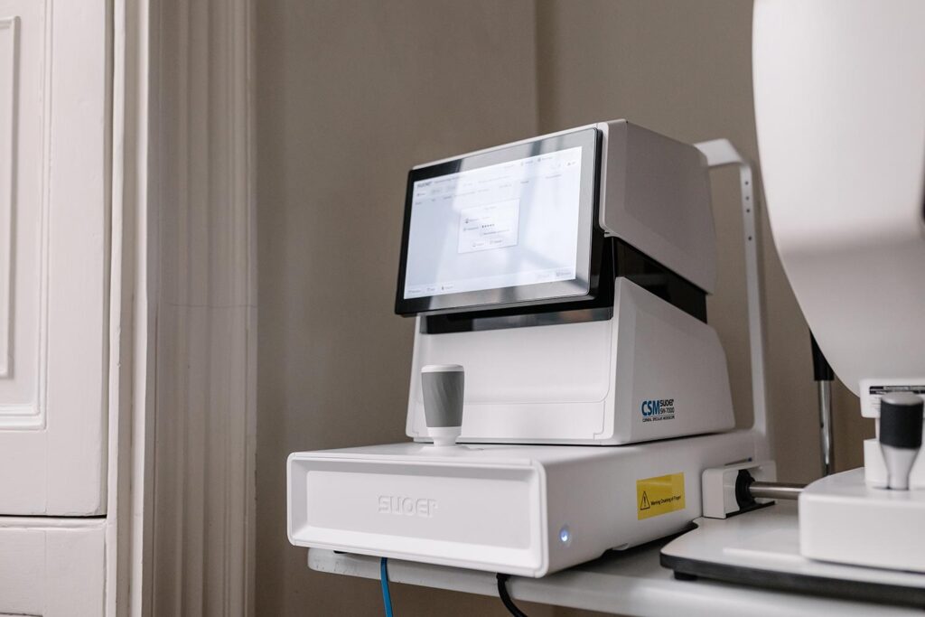
Suoer SW-7000 Specular Microscope

White clinical instrument with a touchscreen monitor mounted on top, placed on a desk in a lab/clinic setting.

Examines the health and density of the corneal endothelial cells, helping confirm your cornea is healthy before laser eye surgery or lens replacement procedures.

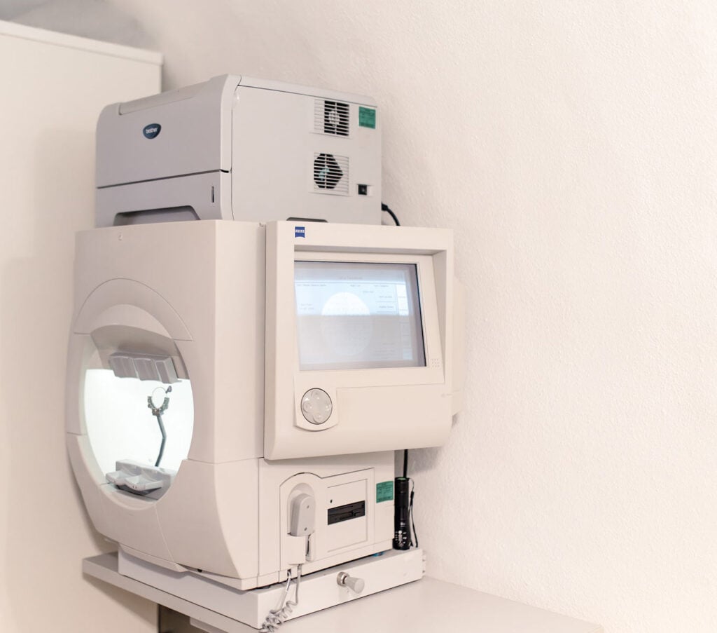
ZEISS Humphrey Visual Fields Analyzer

MRI scanner with a circular opening and patient table ready for a scan.

Assesses central and peripheral vision for subtle areas of sight loss, supporting the detection and monitoring of glaucoma and other eye conditions.

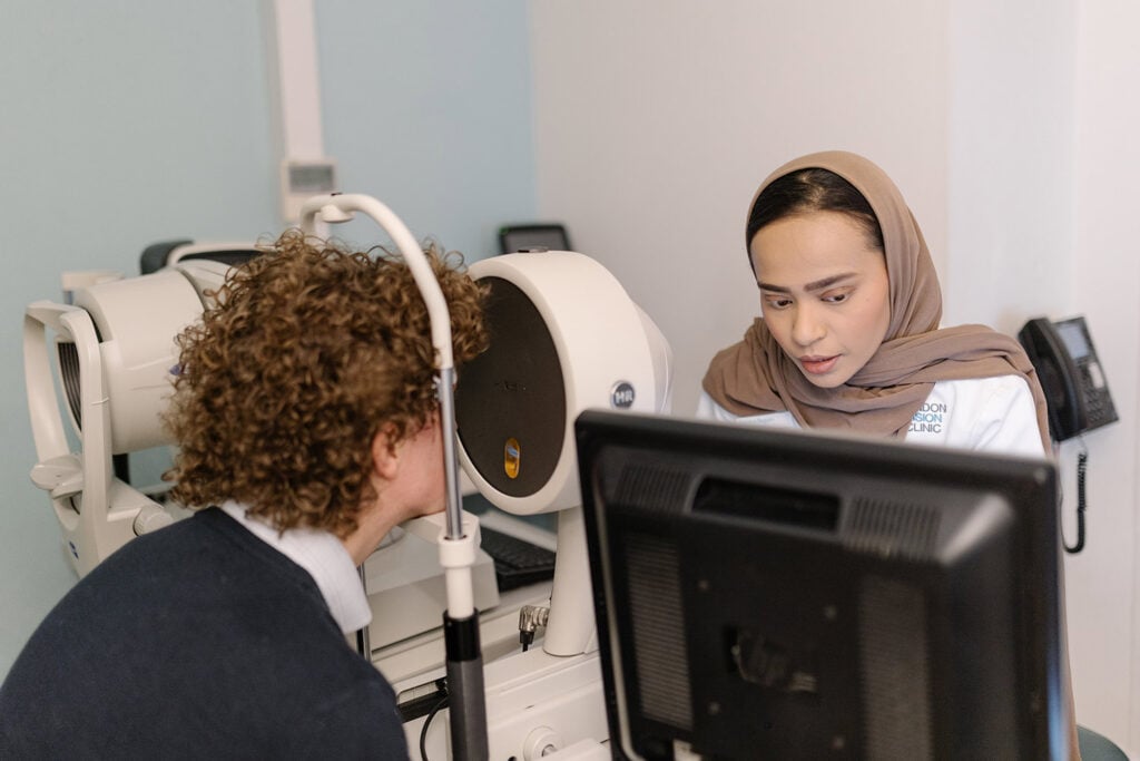
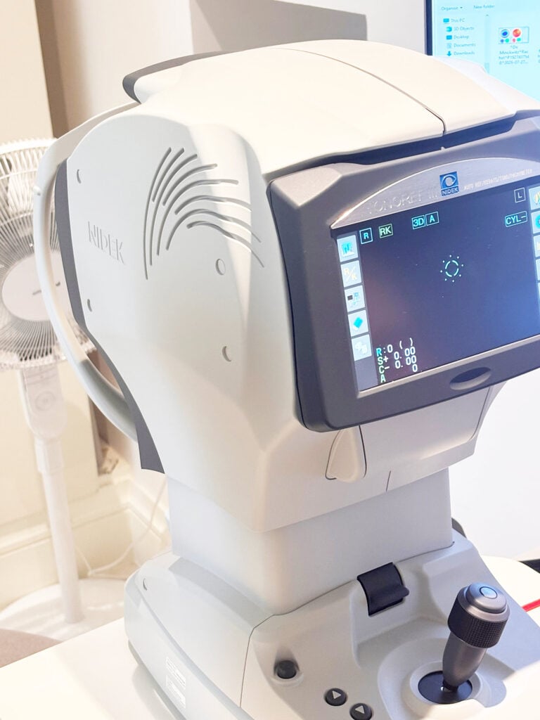
NIDEK Tonoref III
Autorefractor

Ophthalmic diagnostic machine (autorefractor/keratometer) with joystick and touchscreen for eye measurements, labeled NIDEK.

Measures your glasses prescription, the shape of the front of your eye and the pressure inside your eyes. These  measurements help us assess your suitability for laser or lens surgery.

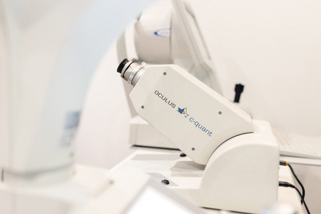
C-Quant Straylight Meter

CQuant

Measures how light scatters inside the eye, helping assess glare and visual quality before laser eye surgery, cataract surgery or lens replacement procedures.

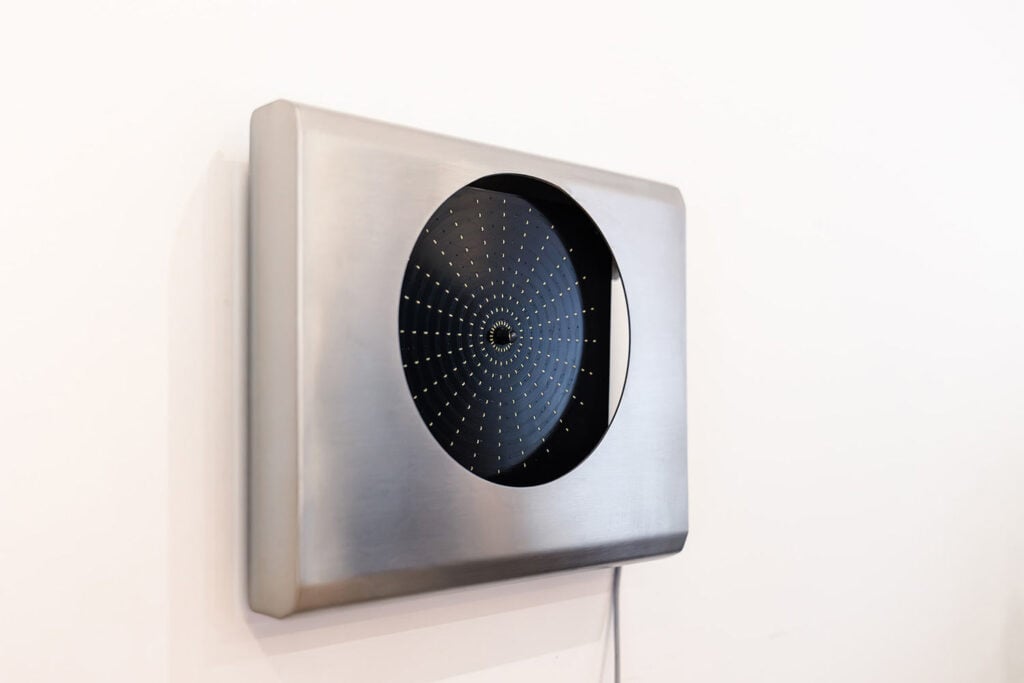
Light Disturbance Analyzer

Close-up of a brushed stainless steel panel with a circular black perforated dial.

Measures halos, glare and other light disturbances, helping assess night vision quality before and after laser eye surgery or lens replacement procedures.

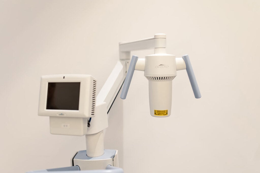
Avedro KXL Cross-Linking

Medical imaging device with two angled arms and a sensor head, mounted on stands in a clinical room (dental radiography setup).

Strengthens weakened corneal tissue using UV light and riboflavin, helping stabilise keratoconus and reduce the risk of further corneal deterioration.

CSO Osiris Wavefront Aberrometer

The CSO Wavefront Aberrometer measures the path light takes as it enters the eye. The Osiris has a unique design that enables it to measure aberrations with a resolution of 45,000 points; 400 times more than the next closest device on the market! This extremely high-resolution wavefront helps to build a detailed picture or map of the eye. This technology is used to build a custom-programmed treatment for each patient and each eye.

Book A Callback