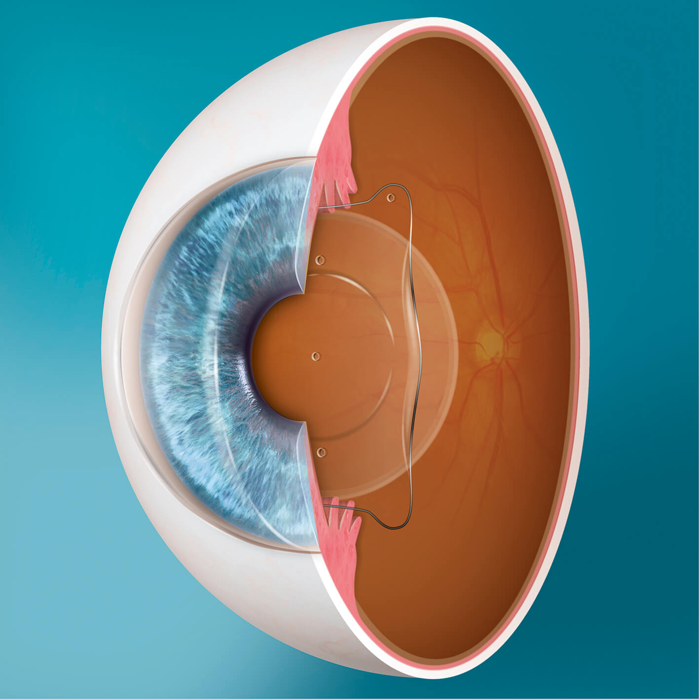
Implantable Collamer Lens (ICL) surgery is the most commonly performed type of Phakic Intraocular Lens Implantation.
This procedure involves implanting a small, thin lens behind the iris and in front of the eye’s natural crystalline lens. ICLs are extremely versatile and can be used to treat almost all prescriptions. ICLs are also available in a range of sizes.

In the UK, ICL Surgery can be used to correct short-sightedness from -0.50 D to -18.00 D, long-sightedness from +0.50 D to +10.00 D, and astigmatism up to 6.00 D.
While earlier types of ICLs were previously linked to an increased risk of cataract development, more modern models (including the EVO Visian ICL) feature a tiny hole in the centre of the lens. This hole – known as the “aquaport” – allows for better flow of aqueous and nutrients around the eye’s natural lens. This helps to mitigate much of the risk of early cataract formation.
The EVO Visian ICL is commonly used in the correction of myopia (short-sightedness); however, they are not used in patients with hyperopia (long-sightedness). Long-sighted patients may instead require a short laser procedure before their ICL surgery to create a small hole in the iris for fluid to flow through.
To date, over one million ICLs have been implanted worldwide.
Eligibility for ICL Surgery
Your suitability for ICL surgery will be determined through an extensive consultation process. Various tests and examinations are performed to assess the health and various measurements of the eye, including the vitreous and retina. For example, state-of-the-art wide-field imaging to check for certain retinal pathologies that could affect the success rate of ICL surgery.
It may also be necessary for a retinal specialist to evaluate the stability and health of your retinas. Some patients may need to undergo treatment for asymptomatic retinal holes or tears before being approved for ICL surgery.
The ICL Procedure
ICL Surgery is usually performed on one eye at a time, with around 1-3 days between each procedure. Each treatment takes around 10 minutes and is followed by a final check, including an eye pressure and lens position review.
After the Procedure
Most patients will be able to return to many normal activities the day after their procedure; however, there will be some restrictions that will be explained by your surgeon. Generally, swimming, intense physical activities, and dusty or dirty environments should be avoided for at least a few days.
ICL patients will typically be advised to apply lubricating eye drops for 2-3 weeks following their treatment. This will help to manage any discomfort or dryness associated with the procedure. Some patients may also experience temporary side effects such as glare around light sources, particularly in low light conditions.
It is extremely rare to need to have ICLs removed due to side effects; however, they can be removed without affecting your pre-corrected vision.
Risks and Complications of ICL Surgery
The most common risks of ICL surgery are pupillary block, cataract formation, and glaucoma. However, these complications are still rare and most are associated with the use of incorrectly sized lenses.
The most suitable lens size should be determined based on several measurements taken during a comprehensive pre-operative assessment. For example, exterior measurements of the eye are often used to estimate the dimensions of the inside of the eye. However, this isn’t always the most effective way to determine the most suitable ICL size as exterior measurements do not always correlate sufficiently with the internal structure of the eye.
With the help of the latest technology, it is possible to collect more accurate measurements. The Artemis Insight 1000 VHF digital ultrasound scanner allows surgeons to directly measure the area inside the eye where the lens will sit. This makes for more accurate ICL sizing and a reduced risk of complications.
If you are considering ICL Surgery, ensuring your provider has access to the latest technology can minimise the chance of experiencing complications after your treatment.

